Nutrition Intervention for Alcoholism: Examining the Consequences and Benefits of Addressing Deficiencies in Recovery
Alcohol addiction can lead to a host of nutrition-related health consequences, including malnutrition, vitamin deficiencies, and damage to the digestive system. Nutrition therapy plays an important role in helping addicts recover.
The Effects of Protein Supplement Timing on Weight Loss
Few understand the complexities surrounding protein intake timing and the impact on weight loss. The effects of protein supplement timing can vary depending on the type of protein supplement you choose.
Nutrient Depletion of U.S. Farmlands and Soil: A Critical Review
Are Americans suffering from nutrient deficiencies due to poor soil? Humans must consume specific amounts of vital vitamins and minerals daily. Crops acquire these nutrients from the soil they grow in, and animals from the crops they eat.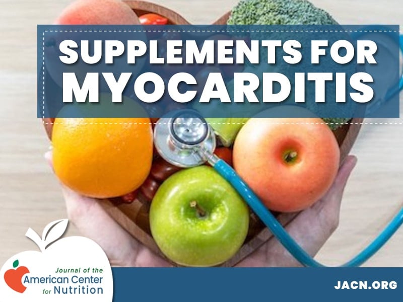
Efficacy of Natural Supplements for Treatment of Viral Myocarditis
While there is no cure for myocarditis, modern clinical studies support the effectiveness of taking natural supplements for treatment of viral myocarditis. Our meta analysis focuses on several of those compounds for efficacy.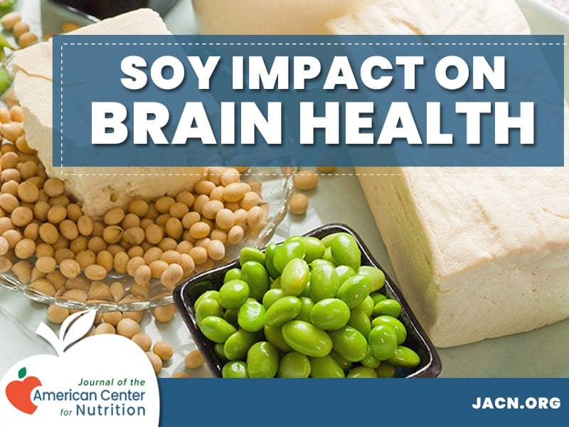
The Impact of Soy Consumption on Brain Health – A Meta Analysis
Various studies have explored the effects of soy consumption on brain health to understand its effects on cognitive function, memory, and existing degenerative diseases. This meta-analysis is focused on reviewing those studies.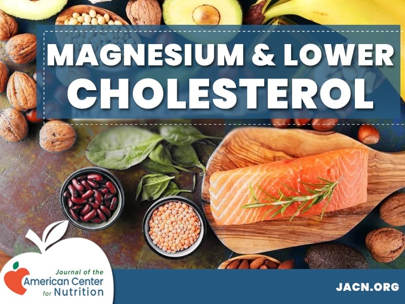
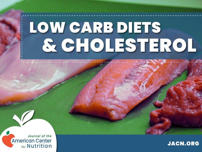
Effects of Magnesium Supplementation on Cholesterol Levels
Evidence suggests an inverse relationship exists between magnesium intake and LDL. Studies show that supplementing magnesium or consuming it in your diet provides numerous health benefits.
Comparison of Whey vs. Plant-Based Protein in Weight Loss and Muscle Building
Healthy adult bodies require protein to function, and many people boost their dietary protein intake with protein powders and supplements to help them reach their fitness goals. The ideal protein source for different people depends on their fitness goals and dietary needs.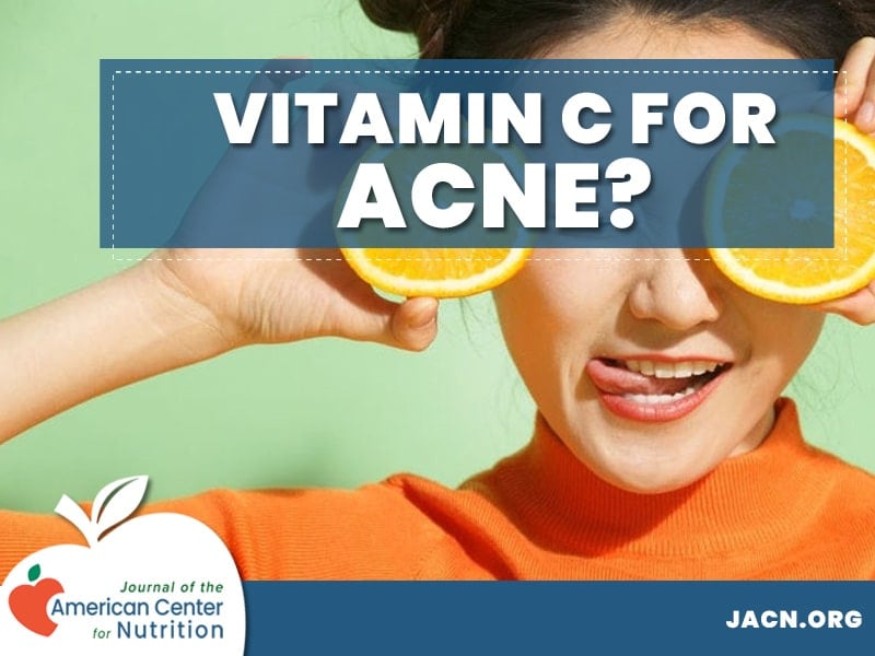
Vitamin C as an Acne Treatment and The Role of Antioxidants in Skin Health
Much research has been done on vitamin c as an acne treatment. Several clinical studies have shown that both topical and oral vitamin c use have a measurable impact on acne through multiple pathways. Acne is a common inflammatory skin condition caused by blocked hair follicles.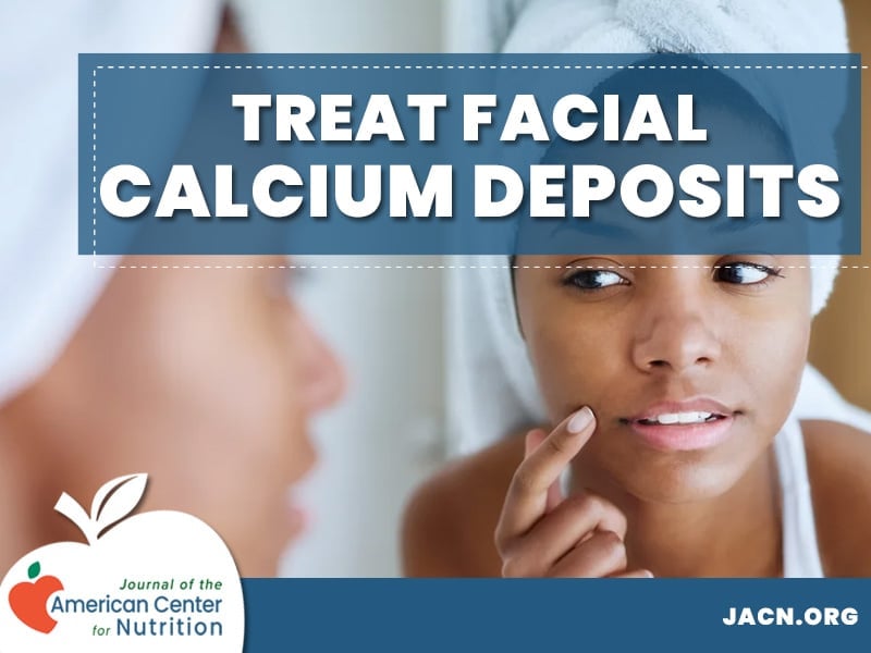
Facial Calcinosis Cutis: Causes, Symptoms, and Treatments
Calcium deposits under the skin are especially bothersome when they appear on the face. Facial calcinosis cutis can be caused by several conditions and we look at various studies for causes and potential cures.