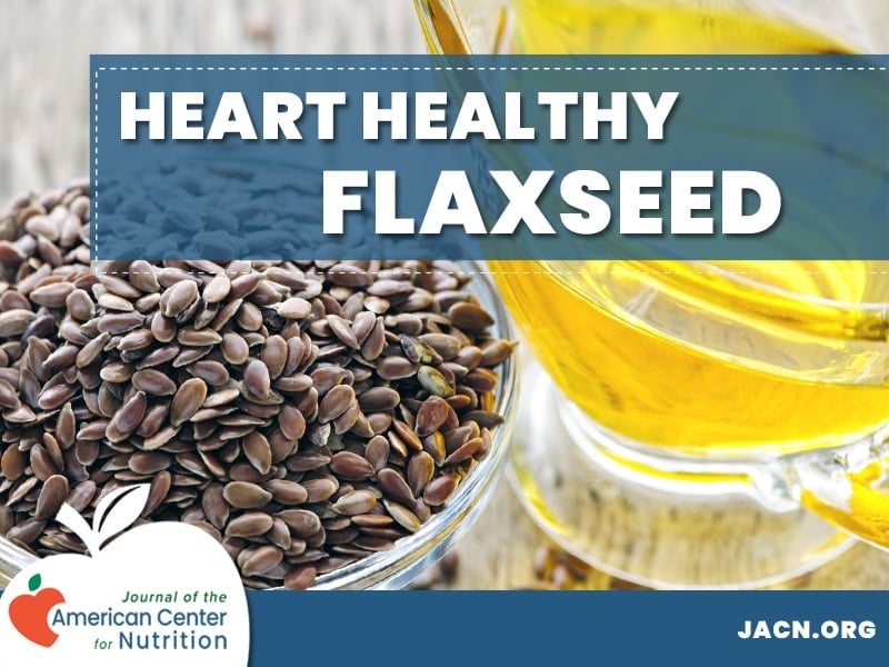
|
|
||||||||
Division of General Internal Medicine (P.O.S., L.T.B.)
Institute for Translational Medicine and Therapeutics (P.O.S., L.T.B., D.J.R., S.B.)
Center for Clinical Epidemiology and Biostatistics (P.O.S., J.B., J.C.)
General Clinical Research Center, University of Pennsylvania School of Medicine, Philadelphia, Pennsylvania, Research Center on Aging, University of Sherbrooke, Quebec, CANADA (S.C.C.)
Address correspondence to: Philippe Szapary MD, MSCE, Cardiovascular Risk Intervention Program, University of Pennsylvania Health System, Philadelphia Heart Institute, 39th and Market streets, suite 2A, Philadelphia, PA 19104. E-mail: philippe.szapary{at}uphs.upenn.edu
| ABSTRACT |
|---|
|
|
|---|
Methods: Sixty-two men and post-menopausal women with pre-study low density lipoprotein cholesterol (LDL-C) between 130 and 200 mg/dl were randomized to 40g/day of ground flaxseed-containing baked products or matching wheat bran products for 10 weeks while following a low fat, low cholesterol diet. Fasting lipoproteins, measures of insulin resistance, inflammation, oxidative stress, and safety were assessed at 0, 5 and 10 weeks.
Results: Flaxseed was well-tolerated, and increased serum levels of ALA (p < 0.001). Compared to wheat, flaxseed significantly reduced LDL-C at 5 weeks (–13%, p < 0.005), but not at 10 weeks (–7%, p = 0.07). Flaxseed reduced lipoprotein a (Lp[a]) by a net of 14% (p = 0.02), and reduced the homeostatic model assessment of insulin resistance (HOMA-IR) index by 23.7% (p = 0.03) compared to wheat at 10 weeks, but did not affect markers of inflammation (IL-6, Hs-CRP) or oxidative stress (ox LDL, urinary isoprostanes) at any time points. In men, flaxseed reduced HDL-C concentrations by a net of 16% (p = 0.03) and 9% (p = 0.05) at 5 and 10 weeks, respectively.
Conclusions: Ground flaxseed has a modest but short lived LDL-C lowering effect, yet reduces Lp(a) and improves insulin sensitivity in hyperlipidemic adults. The HDL-C lowering effect of flaxseed in men warrants additional study.
Key words: flaxseed, hypercholesterolemia, lipoproteins, insulin resistance, inflammation, oxidative stress
| INTRODUCTION |
|---|
|
|
|---|
| MATERIALS AND METHODS |
|---|
|
|
|---|
2.0 mg/dL, and use of any dietary supplements (excluding a daily multivitamin, calcium or vitamin D), hormone replacement therapy, or lipid lowering medications. The protocol was approved by both the General Clinical Research Center (GCRC) and the Institutional Review Board (IRB) at the University of Pennsylvania. The study was fully explained to all volunteers, who provided written, informed consent.
Treatment Products
Whole yellow omega flaxseed (North Dakota Oilseed Council, Flax Section, Bismarck, ND) and durum wheat bran used during the entire study came from the same lots and were ground to powder in ND. Both ground products were stored in opaque containers for the entire study in –20 degree C freezers in the GCRC until they were prepared for consumption. Twenty g of ground flaxseed or wheat bran was baked into 1 of 3 breads or muffins designed and pre-tested to have similar appearance, taste and texture. All products were isocaloric and did not differ significantly in macronutrient or total dietary fiber content (Table 1).
|
38 as well as meeting LDL-C entry criteria, were randomized in a 1:1 double blind fashion to flaxseed or matching wheat control products, stratified by gender. Subjects were instructed to consume one test product in the morning and one in the evening daily for 10 weeks. Test products were prepared in the GCRC 1 day prior to the day subjects were scheduled to receive test agents. Subjects came to the GCRC to pick up test agents at baseline, and weeks 2, and 5. On weeks 4, 7, and 9, frozen test products were sent with an ice pack to the subject via over night mail where subjects were instructed to immediately place products in the refrigerator and store until consumption. Subjects were evaluated at weeks 0, 5 and 10 in the morning after a 12 h fast. At these visits, blood and urine were obtained for changes in safety parameters (chemistry and complete blood count); lipoproteins; and measures of oxidative stress, insulin sensitivity, inflammatory markers and serum fatty acids. Subjects were questioned about adverse events as well as monitored for changes in weight and blood pressure. Systolic blood pressure and diastolic blood pressure were measured with an automatic electronic sphygmomanometer, in the sitting position after resting for at least 5 minutes. Body weight was measured on a calibrated scale (Tronix digital scale, Carol Stream, IL) in light clothes with shoes off to the nearest 0.1 kg.
Serum Fatty Acids
Serum fatty acids were analyzed from EDTA plasma collected from venous blood which was centrifuged at 2400 x g for 20 min at 4° C and then frozen at –70°F until analyzed using a gas chromatography (GC) method as previously described [21]. Individual fatty acids were identified by their retention time compared to that of authentic standards (NuChek Prep, Elysian, MN & Matreya Inc., Pleasant Gap, PA). Individual and combined fatty acids are reported and analyzed as % of total fat. The intra-assay coefficient of variation (CV) for pooled human plasma was 2.5% for linoleic acid.
Lipoproteins
Lipoproteins were analyzed from EDTA plasma collected after a 12 h fast in a CDC-standardized lipid laboratory. Plasma TC, high density lipoprotein cholesterol {HDL-C), and TG were measured enzymatically on a Cobas Fara II autoanalyzer (Roche Diagnostic Systems Inc., NJ, USA) using Sigma reagents (Sigma Chemical Co., MO, USA). LDL-C and very low density lipoprotein cholesterol (VLDL) levels were determined after ultracentrifugation at a density of 1.006 g/ml. Apolipoprotein (Apo) B and ApoA-I were measured using Wako reagents (Wako Chemicals USA Inc., Richmond, VA). Lipoprotein (a) [Lp(a)] was measured using an immunoturbidometric assay (Wako Chemicals USA Inc., Richmond, VA). The intra-assay and inter-assay CV for pooled human plasma were 2.4% and 1.3% for TC, 2.2% and 1.5% for HDL-C, 1.81% and 1.9% for TG, 2.5% and 2.8% for ApoB, 1.2% and 2.6% for ApoA-I, and 4.3% and 16.8% for Lp(a).
Insulin Sensitivity
Insulin sensitivity was estimated using both fasting homeostasis model assessments of insulin resistance (HOMA-IR) index, and the quantitative insulin sensitivity check index (QUICKI). HOMA-IR was defined as [fasting glucose (mmol/L) x fasting insulin (mU/ml)]/22.5 {Matthews, 1985 1309 /id}. QUICKI was defined as 1/[log fasting insulin (mU/ml) + log fasting glucose (mg/dl)] [22]. Insulin was measured from venous blood collected in EDTA and was centrifuged at 3000 x g for 20 min at 4° C (this same procedure was followed for inflammatory markers and serum oxidized LDL). Glucose was measured from serum in our clinical laboratory as described under safety laboratory parameters listed below, while insulin was measured using double antibody human specific radioimmunoassay (Linco Research Inc., St Charles, MO). The intra-assay and inter-assay CV for human plasma for insulin was 15% and 9.8% and 0.7% and 1.4%, respectively for glucose.
Inflammatory Markers
High sensitivity C reactive protein (hs-CRP) was measured with an ultra high-sensitivity latex turbidimetric immunoassay (Wako Chemicals USA Inc., Richmond, VA) on a Cobas Fara II analyzer (Roche Diagnostics, Indianapolis). Plasma levels of interleukin-6 (IL-6), were measured using commercially available enzyme-immunoassays (ELISAs) according to the manufacturer's guidelines (R+D Systems, Minneapolis, MN). The intra-assay and inter-assay CV for human plasma for hs-CRP was 3.7% and 2.8% and 8.7% and 19.9%, for IL-6. Blood was collected from venous blood in EDTA for both assays.
Oxidative Stress
Oxidative stress was by total concentration of oxidized LDL (oxLDL) from serum as measured by a commercial sandwich ELISA assay using a murine monoclonal antibody (mAb-4E6) (Mercodia Inc., Metuchen NJ) as well as urinary isoprostane 8,12-iso-iPF2
-VI as measured by liquid chromatography/tandem mass spectrometry (LC/MS/MS). The levels of 8,12-iso-iPF2
-VI were normalized to urinary creatinine (Cr) levels and reported as ng 8,12-iso-iPF2a-VI/ mg Cr. The intra-assay and inter-assay CV for human urine were 0.1% and 1.1% for 8,12-iso-iPF2
-VI. The intra-assay and inter-assay CV for pooled human serum were 35.6% and 3.2%, respectively, for oxLDL.
Safety Laboratory Parameters
A standard chemistry panel was performed on serum and centrifuged at 3500 x g for 10 minutes and analyzed on a Vitros 950 analyzer. A complete blood count was performed with a coulter STKS CBC counter from EDTA plasma.
Adherence and Masking
Adherence to test products was judged by self-reported treatment diaries. Masking was assessed by questionnaire, which forced a guess as to what treatment subjects believed they were receiving followed by a question on how sure subjects were of their guess. Adherence with the low-fat and cholesterol diet was assessed by 3-day diet records (2 week days and 1 weekend day, analyzed using Nutrition Data Systems for Research software, version 4.05, University of Minnesota) and MEDFICTS obtained at baseline, 5 and 10 weeks.
Outcomes and Sample Size Calculations
The primary endpoint was percent change from baseline in directly measured LDL-C. This was calculated as [(Week 5 or week 10 LDL - baseline LDL)/baseline LDL] X 100. Secondary endpoints included: percent changes in TC, HDL-C, VLDL-C, TG, Apo AI and B, and Lp(a); changes in HOMA-IR and QUICKI; hs-CRP and IL-6; urinary 8,12-iso-iPF2
-VI /mg Cr and serum oxLDL, and safety laboratory tests. Based on published data, we anticipated net reductions in LDL-C of 8% between the flax and wheat groups at both 5 and 10 weeks. Accounting for a 15% estimated loss to follow up, we estimated that a sample size of 30 per group would provide at least 80% power to detect this difference between the two groups, using a two-tailed alpha of 0.05 and an estimated within group standard deviation of 10%.
Statistical Analysis
The primary analysis was by intent to treat (ITT) using data from all 62 subjects meeting study criteria who were randomized (Fig. 1). Two secondary analyses were performed for all lipid variables including a completers analysis and an adherence analysis. The completers analysis only used those values for subjects who had completed all study visits (n = 58). The adherence analysis included subjects who self-reported eating 80% or more of the study food as judged by treatment diaries (n = 49). Exploratory analyses of flaxseed's effect in patients with hypertension were also performed. While these were not pre-specified analyses, this subgroup represents a higher risk group of patients in whom flaxseed may be more effective. We defined hypertension as self-reported history or taking at least one antihypertensive medication.
|
| RESULTS |
|---|
|
|
|---|
|
|
|
|
80%, 1 person in flax vs. 2 in wheat were non-compliant at week 5 (p = 0.52), while 5 in the flax vs. 2 in the wheat were non-compliant at week 10 (p = 0.21). Data on adherence were missing in 5 subjects (2 in flax and 3 in wheat), which were excluded from the adherence analysis. By 10 weeks, 90% of flax and 77% of wheat assigned subjects believed they had received flaxseed during the study (p = 0.3).
ApoB-Containing Lipoproteins
At time of randomization, there were no differences in any lipoproteins (Table 6). After 5 weeks of treatment, flaxseed significantly reduced TC, LDL-C, and ApoB when compared to the wheat group. These findings at 5 weeks were robust and did not change in the completers or adherence analyses. There was no treatment by gender interaction in terms of ApoB and LDL-C. By 10 weeks, there was no difference in LDL-C between treatment groups in the ITT analysis (Table 6). However, in the adherence analysis, the difference in LDL-C of 9% attained statistical significance (p = 0.05).
|
ApoA-I Containing Lipoproteins
There were no differences in baseline HDL-C among males or females between the treatment groups. We found a significant treatment by gender interaction with HDL-C, but not apo AI, that was present at both 5 and 10 weeks. Thus, the HDL-C is separated by gender in Table 6. Starting at 5 weeks and continuing until the end of treatment, flaxseed significantly reduced HDL-C in men, with no effect in women. Adding baseline HDL-C, weight, physical activity, dietary intake of total or saturated fat, alcohol, or HOMA-IR to a multivariable linear regression model did not change the effect of flaxseed on HDL-C in males. Changes in HDL-C levels in men were modestly negatively correlated to serum ALA at 5 weeks (r = –0.42, p = 0.05) and 10 weeks (r = –0.50, p = 0.02).
Insulin Sensitivity
There was no change in body weight at the end of the treatment period compared to baseline in either group (flax, 79 ± 15 and 79 ± 15 kg at weeks 0 and 10; wheat, 78 ± 15 and 77 ± 15kg at weeks 0 and 10, p value between groups = 0.16). There was no significant change in glucose or insulin in either treatment group at 5 (data not shown) or 10 weeks (Table 4). However, both HOMA-IR and QUICKI indices suggest that flaxseed significantly improved insulin sensitivity when compared to wheat at 10 weeks. Restricting our analysis to most adherent patients and adjusting for BMI only strengthened the flaxseed effect [logHOMA-IR (p = 0.02); QUICKI (p = 0.03)].
Markers of Inflammation and Oxidative Stress
There were no significant changes in hs-CRP at 5 or 10 weeks (Table 4). In an exploratory analysis, when data were dichotomized by favorable response status (subjects whose week 10 hs-CRP was reduced vs. those whose hs-CRP did not change or increased), 18/29 (62%) of subjects given flaxseed responded favorably compared to 11/30 (37%) wheat-treated subjects (p = 0.04). In an exploratory analysis, flaxseed had an anti-inflammatory effect in patients with hypertension (n = 19), reducing hs-CRP from 1.40 to 1.13 mg/L (–19%) compared to an increase from 1.22 to 1.30 mg/L (+9%) in the wheat group (p = 0.02). The change in hs-CRP was negatively correlated to the changes in serum levels of ALA (r = –0.41, p = 0.001). There were no changes in IL-6 levels at either 5 or 10 weeks. Flaxseed did not affect serum oxidized LDL or urinary isoprostane 8,12-iso-iPF2
-VI measures at 5 or 10 weeks (Table 4).
Safety and Tolerability
Forty-one subjects experienced a total of 40 adverse events (AEs), with 20 in each group (p = 0.73). There were 3 serious AEs, all for hospitalizations (intestinal bowel obstruction, cataract surgery, and removal of a thyroid nodule), with the latter two judged as not related and the bowel obstruction judged as possibly related. This subject received flaxseed, which was discontinued at time of notification of hospitalization. The most common reported AEs included diarrhea (19% subjects), flatulence (15% subjects), and headache (5%). There were a total of 13 gastrointestinal-related AEs (diarrhea, constipation, flatulence, nausea, vomiting, dyspepsia, and/or abdominal distension) in the flaxseed group and 8 in the wheat group (p = 0.13).
There were no differences between groups on any component of a complete blood count. Flax reduced aspartate aminotransferase (AST) from a mean 29.9 ± 12.9 U/L to 26.0 ± 10.1 U/L (–3.9 U/L) while wheat slightly increased AST from 27.6 ± 12.6 U/L to 28.5 ± 6.7 U/L (+0.84 U/L) (p = 0.01). Similarly, alanine aminotransferase (ALT) went from a mean of 30.8 ± 15.7 U/L to 26.4 ± 8.4 U/L (– 4.4 U/L) in flax with a slight increase in wheat from 29.7 ± 12.6 U/L to 30.5 ± 6.7 U/L (+0.58 U/L) (p = 0.008). ALT levels at 10 weeks were positively correlated to HOMA-IR (r = 0.35; p = 0.005).
| DISCUSSION |
|---|
The significant reduction in Lp(a) has been reported previously in post-menopausal women [13] but ours is the first to show this in men. Lp(a) is relatively uninfluenced by diet and other lifestyle factors [25]; however, the clinical significance of this finding is uncertain.
We report for the first time that flaxseed significantly lowered HDL-C in men, but not women. Of the published RCTs using flaxseed, 3 have shown reductions in HDL-C ranging from 1 to 7%, but these changes were only significant in a single large trial of postmenopausal women in which 40g of flaxseed reduced HDL-C by a net of 4.6% (p = 0.03) at 1 year compared to wheat germ[19]. The other studies included either no [13,16,17] or few (12,15,24] men. We found that change in plasma ALA levels was modestly negatively correlated to changes in HDL-C, suggesting dietary ALA levels may contribute to this effect. This negative correlation between ALA and HDL-C is in keeping with findings from 2 RCTs [26,27]. Wilkinson et al randomized 57 men to a diet either enriched with ALA from flaxseed oil (19 g ALA/d), linoleic acid (LA) from sunflower oil, or sunflower oil with fish oil for 12 weeks. Plasma HDL-C was reduced by 10.5% in the flax oil group compared to a reduction of 5.6%, and an increase of 3% in the sunflower and sunflower + fish oil groups, respectively (p = 0.009). Another feeding study compared an average American diet, a high ALA diet, and a high LA diet [27] supplying 36% energy as total fat and found a significant reduction in HDL-C of
6% in 23 subjects (20 of whom were male) following the high ALA diet compared to the average American diet. The potential mechanism of an ALA-induced reduction in HDL-C in men is not known.
Flaxseed improved validated indices of insulin sensitivity. While one previous study showed a reduction in glucose area under the curve (AUC) [11], ours is the first study to report improvements in either HOMA-IR or QUICKI. A study by Cunnane et al. found that both whole flaxseed and isolated flaxseed mucilage (fiber alone) reduced the AUC for glucose during a 2 hour OGTT by 27% in 9 healthy women compared to glucose alone, suggesting the fiber portion of flaxseed is primarily responsible for this hypoglycemic effect [11]. The clinical implication from improvements in insulin sensitivity in the range shown by flaxseed is not clear, but could be construed as reducing the risk of developing T2DM.
Flaxseed was also associated with small but significant reductions in liver transaminases. This has not yet been reported in humans and the significance is unknown, but flaxseed has been shown to reduce hepatosteatosis in both lean and obese rats compared to both casein and soy protein [28]. Changes in AST and ALT were correlated to changes in insulin sensitivity, suggesting a possible link between the two. This association has been seen in a larger dataset where elevations in ALT, even within the normal range, have been independently associated with IR, even after adjusting for BMI, HTN, low HDL-C and elevated hs-CRP [29].
This is the first trial to report the effects of ground flaxseed on serum markers of inflammation. While we did not find an effect of flaxseed on IL-6 or hs-CRP in the overall population, there was a significant reduction in hs-CRP in hypertensive patients, though this was a small subset of the total group. We did however find a significant negative correlation between hs-CRP and serum ALA levels as seen in previous studies using ALA-enriched diets from multiple sources [27,30,31].
Early studies have raised concerns that foods and dietary supplements with high levels of polyunsaturated fatty acids such as fish oils [32] or flaxseed [24] could be pro-oxidant. However, more recent literature suggests that this phenomenon may be the result of insensitive ex vivo assays used to measure oxidative stress [33]. This is the first study to investigate the in vivo effects of flaxseed consumption on F2-isoprostanes, a validated in vivo marker of oxidative stress. Our data suggests 40 g ground flaxseed does not increase oxidative stress.
In conclusion, incorporating 40g (4–5 Tbsp) of milled flaxseed into a low fat, low cholesterol diet is feasible and well-tolerated, and provides a good source of soluble fiber and ALA, two key nutrients associated with reduced risk of diabetes and CHD. Flaxseed initially reduced LDL-C, but effects were not sustained at 10 weeks. Flaxseed reduced HDL-C in men, but not women. Our data also suggests flaxseed improves insulin sensitivity, but does not affect markers of inflammation or oxidative stress. Future, mechanistic studies are needed to clarify the reasons for the HDL-C gender differences, as well as to determine the component(s) responsible for effects reported. In addition, longer clinical trials of 6–12 months duration which include a larger (n > 100) and equal number of both men and women are needed to fully assess the potential of this functional food as a component of a healthy cardioprotective diet.
| ACKNOWLEDGMENTS |
|---|
|
|
|---|
| FOOTNOTES |
|---|
|
|
|---|
Current affiliation for J.A.B.: Johnson & Johnson Pharmaceutical Research and Development, LLC.
Received December 5, 2005. Accepted August 28, 2006.
| REFERENCES |
|---|
|
|
|---|
This article has been cited by other articles:
![]() |
M. V. Potenza and J. I. Mechanick The Metabolic Syndrome: Definition, Global Impact, and Pathophysiology Nutr Clin Pract, October 1, 2009; 24(5): 560 - 577. [Abstract] [Full Text] [PDF] |
||||
![]() |
A. E. Barden, K. D. Croft, T. Durand, A. Guy, M. J. Mueller, and T. A. Mori Flaxseed Oil Supplementation Increases Plasma F1-Phytoprostanes in Healthy Men J. Nutr., October 1, 2009; 139(10): 1890 - 1895. [Abstract] [Full Text] [PDF] |
||||
![]() |
A. Pan, D. Yu, W. Demark-Wahnefried, O. H Franco, and X. Lin Meta-analysis of the effects of flaxseed interventions on blood lipids Am. J. Clinical Nutrition, August 1, 2009; 90(2): 288 - 297. [Abstract] [Full Text] [PDF] |
||||
| ||||||||||||||||||||||||||||||||||||||||||||||||||||||||||||
| HOME | HELP | FEEDBACK | SUBSCRIPTIONS | ARCHIVE | SEARCH | TABLE OF CONTENTS |